Introduction
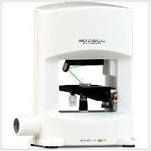
The 3D Cell Explorer is a non-invasive microscope that can image cells & tissues in vitro instantly and in 3D without any stain or label. Through a combination of holography and rotational scanning the system measures precisely the distribution of the physical refractive index (RI) within the cell (in 3D and timelapse).
Thanks to its completely self-adjusting optics, the best imaging results are always guaranteed – whatever the environment is (fundamental patent US 8,937,722 & EU WO 2011/121523).
Technical Specifications

Fetures
5 min of work
Non-invasive
Direct measurement of cell’s properties
Free digital staining
200nm resolution
3D results


Applications
The 3D Cell Explorer is a tool for discovery and we are just at the beginning of exploring all the potential fields of application. There are no boundaries.Thanks to this new technology, we believe that many discoveries on living cells will be done and will extend our understanding of life, diseases and effect of drugs.
Cell imaging conditions enabled by the 3D Cell Explorer:

The 3D Cell Explorer performs 3D and real-time in vitro cell imaging on
A. SINGLE CELL MORPHOLOGY, KINETICS & DYNAMICS
• Death
• Remodeling
• Migration
• Adhesion
• Intracellular trafficking
• ...
B. INTERACTIONS & REACTIONS
• Cell-cell
• Cell-bacteria
• Cell-virus
• Inorganic material
• Stimuli
• Drugs & Toxicity
• Nanomaterial internalization/
trafficking
C. STAIN-FREE HISTOLOGY& HISTOPATHOLOGY
• Tissues morphological analysis
• 3D Tissue characterization
• Cancer diagnosis
• ...
D. STAIN-FREE CYTOPATHOLOGY
Cell nuclear evaluation
• Fast Pap-smear analysis
• Needle aspiration biopsy
• Cancer diagnosis
• Infection diagnosis
• Microbial infections:
parasitic, viral, and/or bacterial
• Reactive changes
• Immune reactions
• Cell aging
• Amyloidosis
E. STAIN-FREE BLOOD CYTOLOGY
Drop of blood based diagnosis of:
Red blood cells count (CBC) (e.g. Anemia)
• White blood cells count (e.g. Mononucleosis)
• Platelets count (e.g. Leukemia, Myeloma)
• Microorganism infections (e.g. Malaria, Chlamydia trachomatis)
• Monitor size and shape of the red blood cells (e.g. Anemia)
• Shape, size, and relative numbers of white blood cells
(e.g. Sickle-cell disease, G6PD defi ciency)
F. CELL’S METRICS
Metrics
Area
• Thickness
• Volume
• Texture/Roughness
• Irregularity
• Activity (moving fromframe to frame)
• Density distribution *histogram of RI
• RI segmentation (d-stains)
• Autoimmune diseases